Ultraviolet Fluorescence in Plants - Light in the Fast Lane
Pulling back the curtain on the secret diversity of wild fluorescence in wild plants
by Jay Vannini
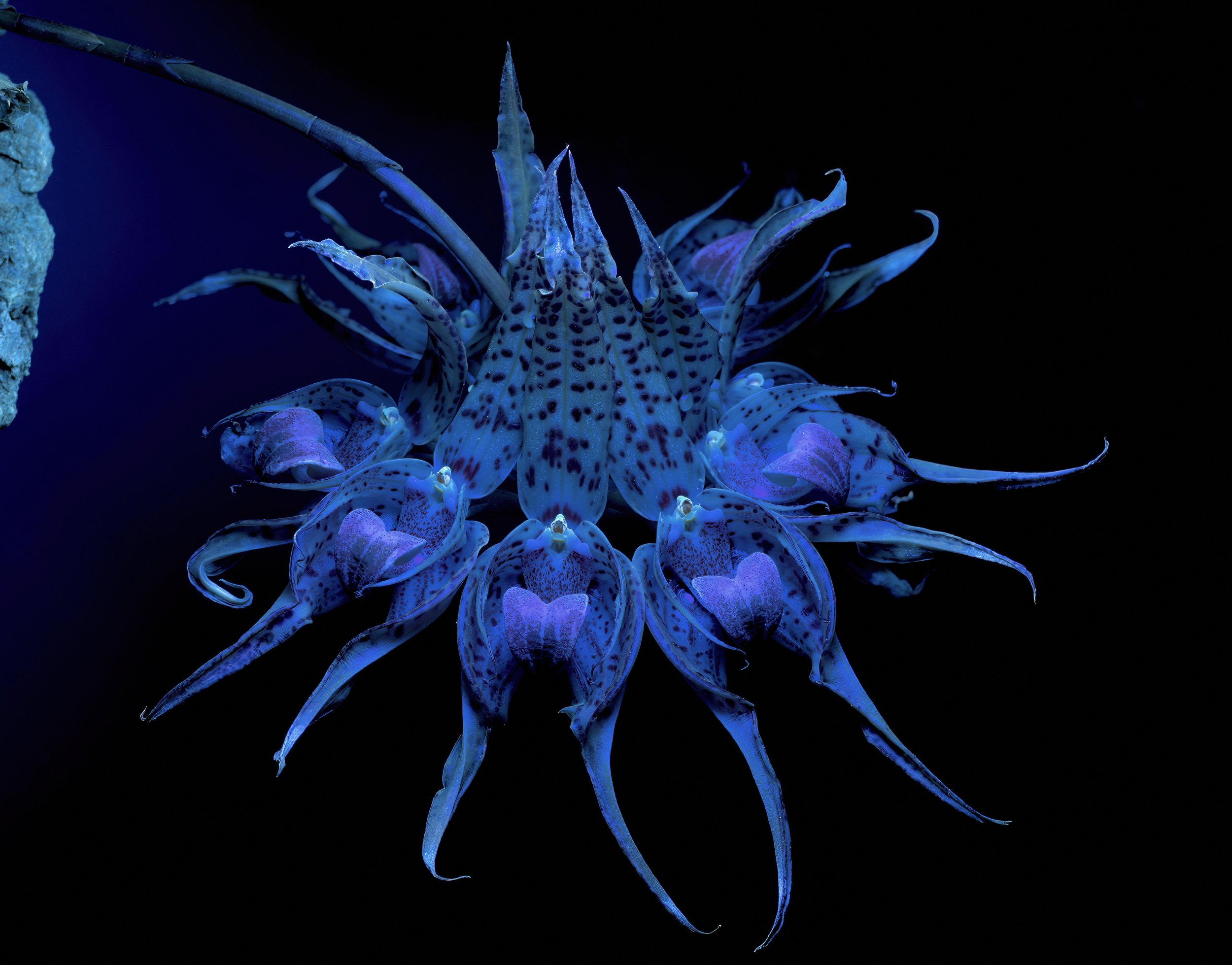
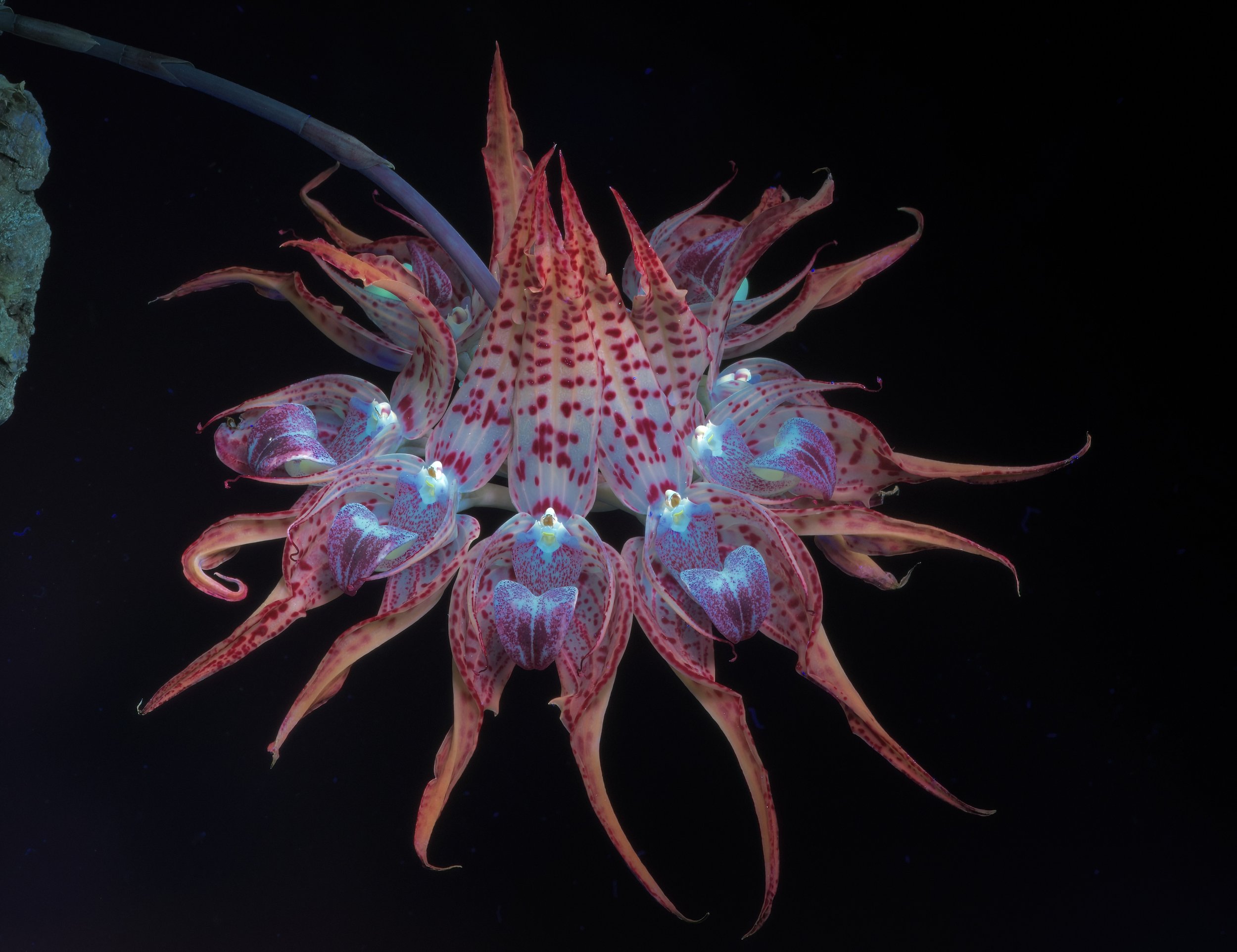
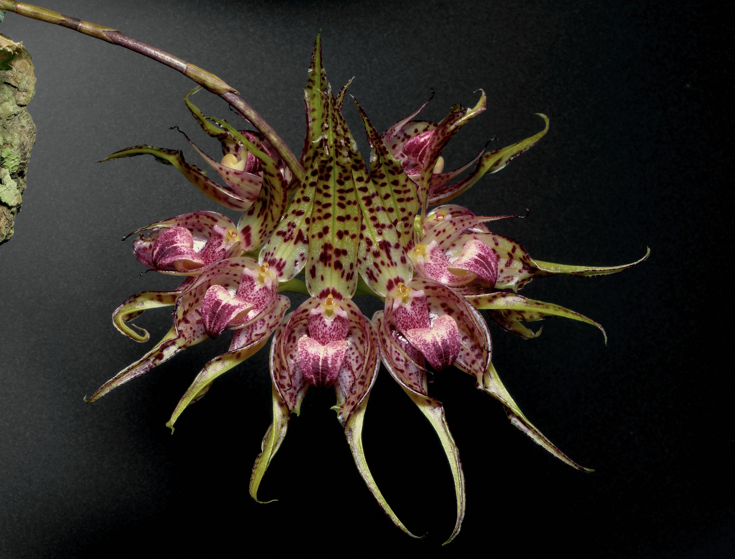
A slideshow of the bizarre inflorescence of a Javan orchid, Bulbophyllum ericssonii, exposed to shortwave ultraviolet (deep blue - 254 nm), longwave ultraviolet (365 nm), and daylight (535 nm) wavelengths. This distinctive flower form was originally described as B. binnedijkii and still circulates in the trade under that name. The decision to place B. binnendijkii and B. virescens into synonymy with B. ericssonii remains controversial among some orchid collectors. Author’s collection. Images: ©Jay Vannini 2023.
Part I: Optics, Plant Pigments & Gallery
In a universe filled with marvels, light is the greatest marvel of all.
From a human perspective, light brings both practical and symbolic remedies to our existence. Light sparks life, feeds us, generates warmth, marks time, brings hope.
We open our eyes to it when we are born and close our eyes to it when we die.
And somewhere between birth and death, every day we interact with light across many spectra. One of these spectra is ultraviolet. And, happily as it turns out, ultraviolet light can stimulate colorful fluorescence in a variety of objects, including naturally occurring ones that curious humans ponder and collect.
Interest in ultraviolet fluorescence in nature is growing rapidly and research into this segment of optics spans both earth and life sciences.
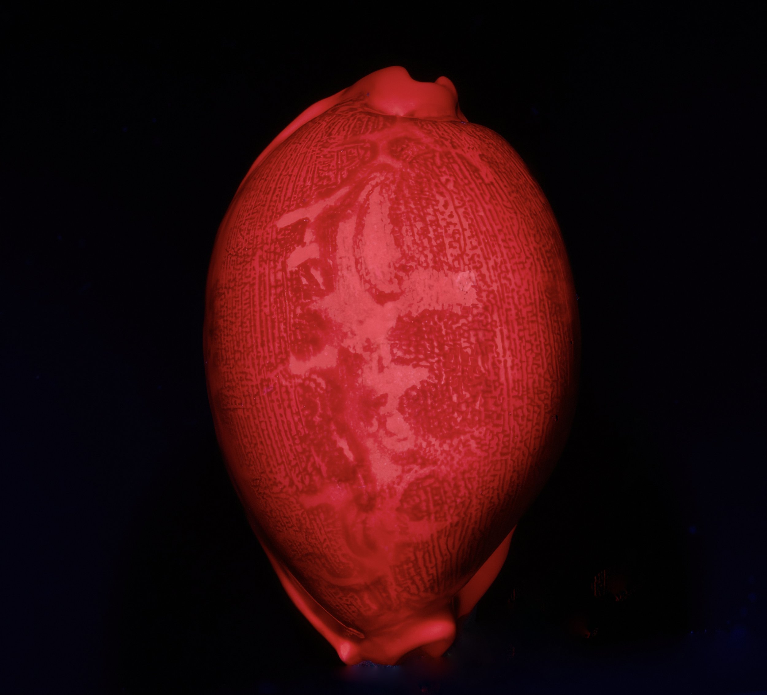
Colorful examples of objects that exhibit UVIVF are found everywhere in nature. While most seashells do not fluoresce under longwave UV, some cowries (Cypraeidae) are famous for it. Shown above, a Geographic Cowrie, Leporicypraea geographica, from Sumbawa Island, Indonesia lit with a 365 nm UV source. Ron Parsons collection. Image: ©Jay Vannini 2023.
Conspicuously fluorescent life forms, especially certain fungi and higher plants, marine invertebrates including corals, anemones, and jellyfishes, together with terrestrial invertebrates such as spiders, scorpions, and centipedes, are among nature’s greatest Easter Eggs (Marshall & Johnsen, 2017). Selectively bred, highly fluorescent freshwater tropical fish (e.g., GloFish®) have been available to home aquarists for two decades. Live stony corals and other fluorescent tropical marine invertebrates are now popular items and routinely displayed under long-wave UV lights in public and home aquaria.
Fluorescent minerals, their collection, and specialized photography are a popular and growing subset of rockhounding and mineral display in public collections. Many mineral species exhibit spectacular fluorescence under mid- and longwave UV lights due to the presence of specific impurities or “activators” contained in their crystals.
My interest in fluorescence revealed under blackout conditions was sparked earlier this year by its conspicuous presence in some minerals and seashells I was photographing, which then led to exploration of its presence and prevalence in my cultivated plant inventories.
As the photos included here will show, Ultraviolet-Induced Visible Fluorescence (UVIVF) in plants is an often colorful and incredibly variable phenomenon and – under controlled conditions – provides live plant collectors with an entirely new perspective from which to view their wards. Differential, distinctive fluorescence on same growth stage leaves on related taxa may also provide botanists with new markers to explore taxonomic relationships within some plant families (pers. obs.). Breeding and genetic engineering to produce daylight fluorescent floral forms are being evaluated (Guerrero-Rubio et al., 2020).
Are sunlight-fluorescent tiger lilies coming soon to a florist near you?
A small bit of requisite physics of optics is required to start off with so that readers understand the “whys” of the dramatic color changes across different light spectra that are shown below, and to understand the terminology used throughout.
No worries for the science-challenged. I have laced this article with plenty of colorful images to satisfy those who found creative excuses to skip their physics classes.
On light
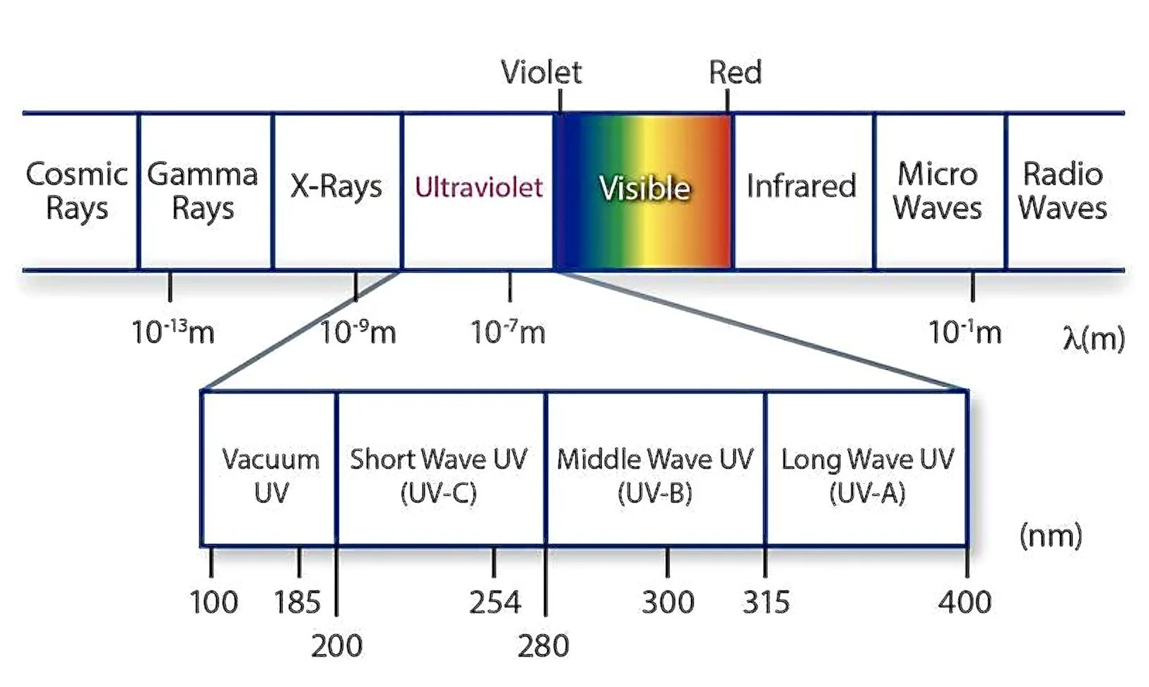
Light is part of the electromagnetic spectrum, a scale that ranges from cosmic and gamma radiation at the lower end to long radio waves at the upper. Light wavelengths are measured in billionths of a meter (expressed as nanometers and abbreviated nm), with visible light, or Photosynthetically Active Radiation (PAR) ranging from ~400 nm (violet) to ~700 nm (red). Within the electromagnetic spectrum, ultraviolet light (UV) ranges from ~10 nm to ~400 nm and is sandwiched between X-Rays on the lowest end (where it is known as extreme UV) and visible light at the upper. Daytime fluorescence in some minerals and organisms is sometimes evident in bright sunlight right around the 400 nm wavelength that is the generally accepted boundary between UV and visible light in most humans.
Extreme and vacuum UV aside, ultraviolet light is further split into three sub bands: UV-C at wavelengths from >200 to 280 nm, UV-B from >280 to 315 nm, and UV-A from >315 to 400 nm; at the upper boundary it becomes visible to most humans. In common usage, UV-C is termed “shortwave UV”, UV-B is “midwave UV”, and UV-A is “longwave UV”. In nature, all UV-C is absorbed by the ozone layer in the stratosphere so does not enter the atmosphere. In controlled settings it is commonly employed in laboratories and potable water processing due to its potent germicidal properties. In limited quantities, midwave UV does make it to the earth’s surface. Prolonged doses of lower-end wavelength UV-B in the atmosphere poses constant risks to humans by causing sunburn (erythema), and via the suppression of the immune system over long-term exposures, sometimes causing skin cancers. Longwave UV is not significantly filtered by the ozone layer. Some UV-A exposure is required to synthesize vitamin D, although again too much exposure is linked to undesirable outcomes such as skin thickening, cancers, and cataract development.
The same animal, floral part, or mineral field under observation will usually fluoresce differently – or not at all – under UV-A, UV-B, and UV-C exposures. From a biologist’s, geologist’s, or photographer’s perspective, since only the latter two UV sub bands occur in nature at all terrestrial elevations and are of principal interest, the use of UV-C lumination in research is primarily of value in exploration the range of colors generated across the UV light spectrum.
Optics, here referring to the branch of physics dedicated to the behavior and properties of light, is a fascinating scientific subject but can be daunting to laypersons. I’ll close with a quote that references the complexities of its more arcane corners by a specialist in this field (Johnsen, 2012):
“…Although a photon (or light wave) has only three properties – frequency, wavelength, and polarization – the long history of light measurement and its association with human vision has left a trail of perplexing and comical units. Only in optics do people still publish papers using units like stilbs, nits, candelas, trolands, and my personal favorite, foot-lamberts. Unless you work in human visual psychophysics, where these units are entrenched, my advice is simple. If you know what these units are, forget them. If they are unfamiliar to you, never learn them…”.
What exactly is fluorescence?
Let’s begin with what it is not.
It is neither the same phenomenon nor a term to be used interchangeably for bioluminescence, chemiluminescence, triboluminescence, iridescence, and phosphorescence (Johnsen, 2012).

California coastal native Moon Jellyfish, Aurelia labiata. These ethereal, short-lived zooplankton-feeding marine invertebrates are bioluminescent due to chemical reactions that occur in their bells. Image: ©Jay Vannini 2023.
Despite common misconceptions that arise from confusion with all the phenomena mentioned in the prior paragraph, fluorescence does not “generate” light. That said, some organisms and minerals that fluoresce vividly under longwave UV may also exhibit bioluminescence (e.g., some soft corals and centipedes), or short-duration phosphorescence (e.g., barites and calcites from certain localities - see image below).
The term originated in the mid 19th century and derives from observations made by noted British physicist Sir George Stokes that fluorspar (calcium fluoride in mineral form, now known as fluorite) glowed under UV light.
Above, twinned images of a strongly fluorescent fluorite specimen from Bergmännish Glück Mine, a silver and cobalt mine in Saxony, Germany that was commercially active in the 18th and early 19th centuries. The fluorite crystals have zoned phantoms visible as darker areas within under both 535 nm daylight and 365 nm longwave UV. The black freckles are pyrite. Interestingly, the two small barite crystals that are fluorescing white in the image on the right also phosphoresce after being exposed to longwave UV, and glow visibly in darkness for almost five seconds after the UV lights have been turned off. Author’s collection. Images: ©Jay Vannini 2023.
Fluorescence is the type of luminescence whereby visible light is generated by some subjects after excitation of their atoms following exposure to normally invisible, low-wave light spectra, primarily ultraviolet (UV), but also X-Ray. It derives from illumination of fluorophores (fluorescent chemicals), that have the capacity to re-emit light at a higher wavelength when exposed to lower wave light spectra.
More specifically, fluorescence is the phenomenon whereby shortwave light in the form of photons is absorbed by a subject in the ground/original orbital state, enters an excited state and shifts to a higher orbital state while exposed to high-energy, shortwave UV photons, and is then immediately re-emitted at a higher wavelength as it returns to the ground state/original orbital after giving off the absorbed energy from UV exposure. Simply put, it is this abrupt change in wavelength that prompts a temporary color change – or fluorescence – under blackout conditions.
Plant pigments in chloroplasts and chromoplasts provide the color to soft plant tissue that are visible to humans in daylight. Most commonly, above-ground soft plant parts appear some shade of green to our eyes in daylight. This is due to chlorophyll pigments in the surface cells of their tissues.
It is widely known among biologists, chemists, and many secondary school science students that chlorophyll is vividly red-fluorescent in a cuvette when illuminated with longwave UV, and that the leaves and reproductive structures of many common plant species will “glow” red or reddish when luminated by a blacklight or longwave UV flashlight. However, as the wavelength of the light emitted by the source decreases, so does red fluorescence in plant parts (pers. obs. - see example below).
Investigation into chlorophyll fluorescence mostly involves chlorophyll a and b, that are often referred to as Chl a and Chl b. Chlorophyll a absorbs violet and orange spectra, while chlorophyll b absorbs primarily in the blue and yellow range. Because chlorophyll in leaves absorbs only minute amounts of green light in the visible spectrum via internal reflections across cells, most leaves appear green in daylight due to reflection rather than absorption. Likewise, because neither absorbs light at the red end of the visible spectra, chlorophyll fluoresces red when exposed to blacklights.
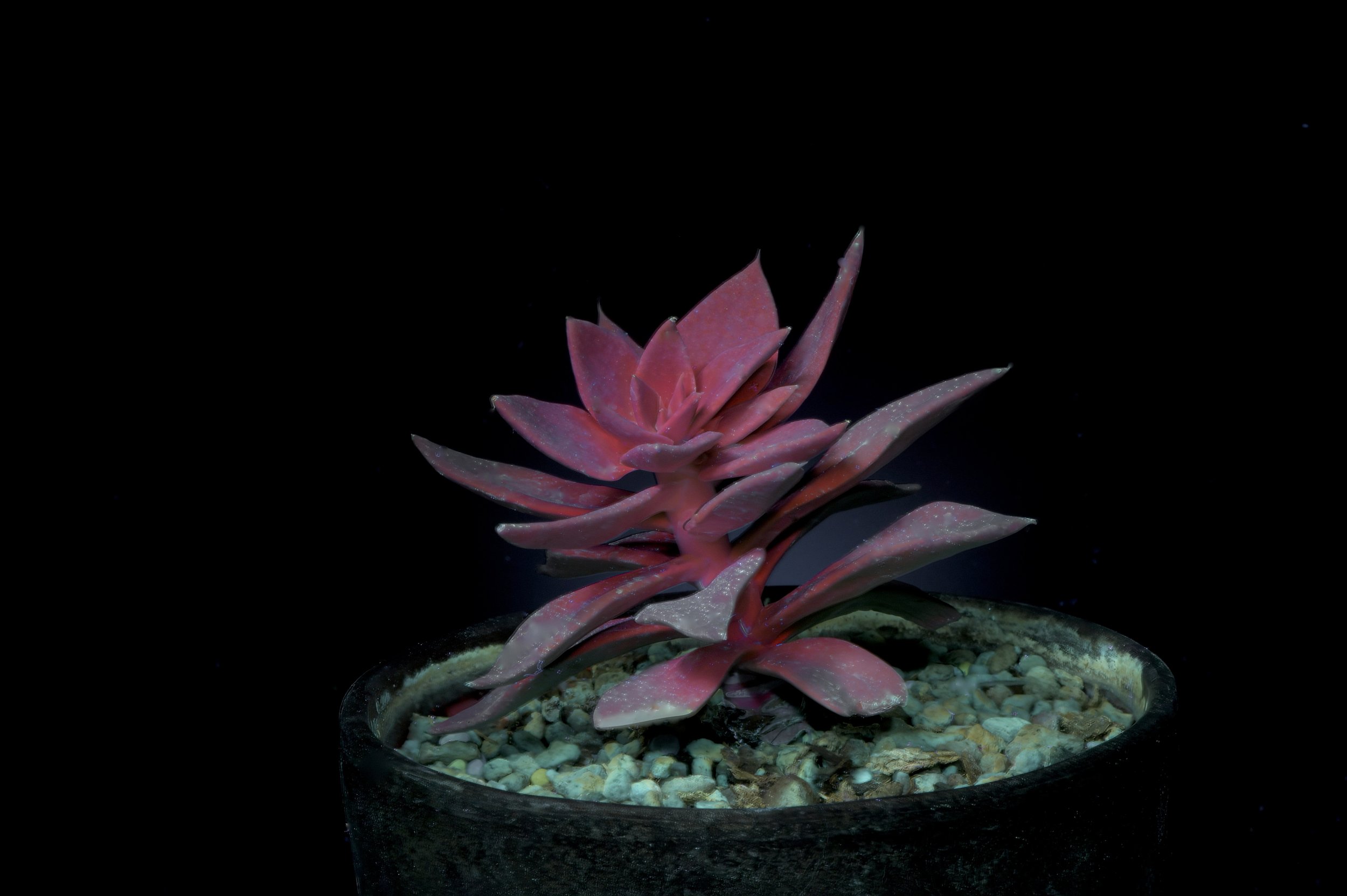

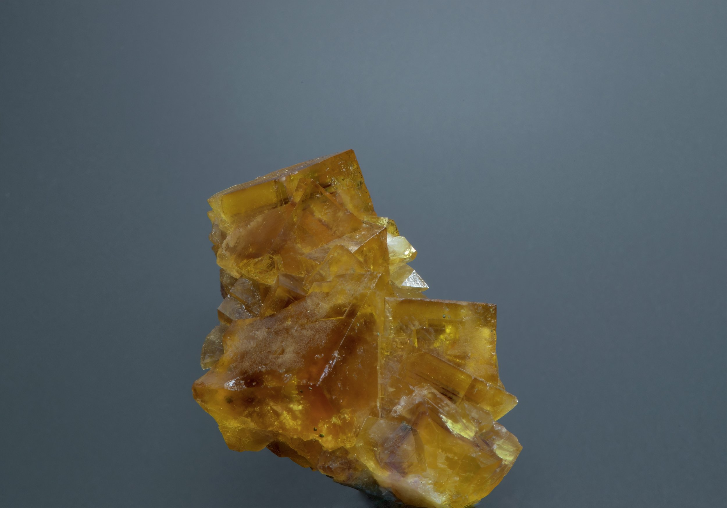
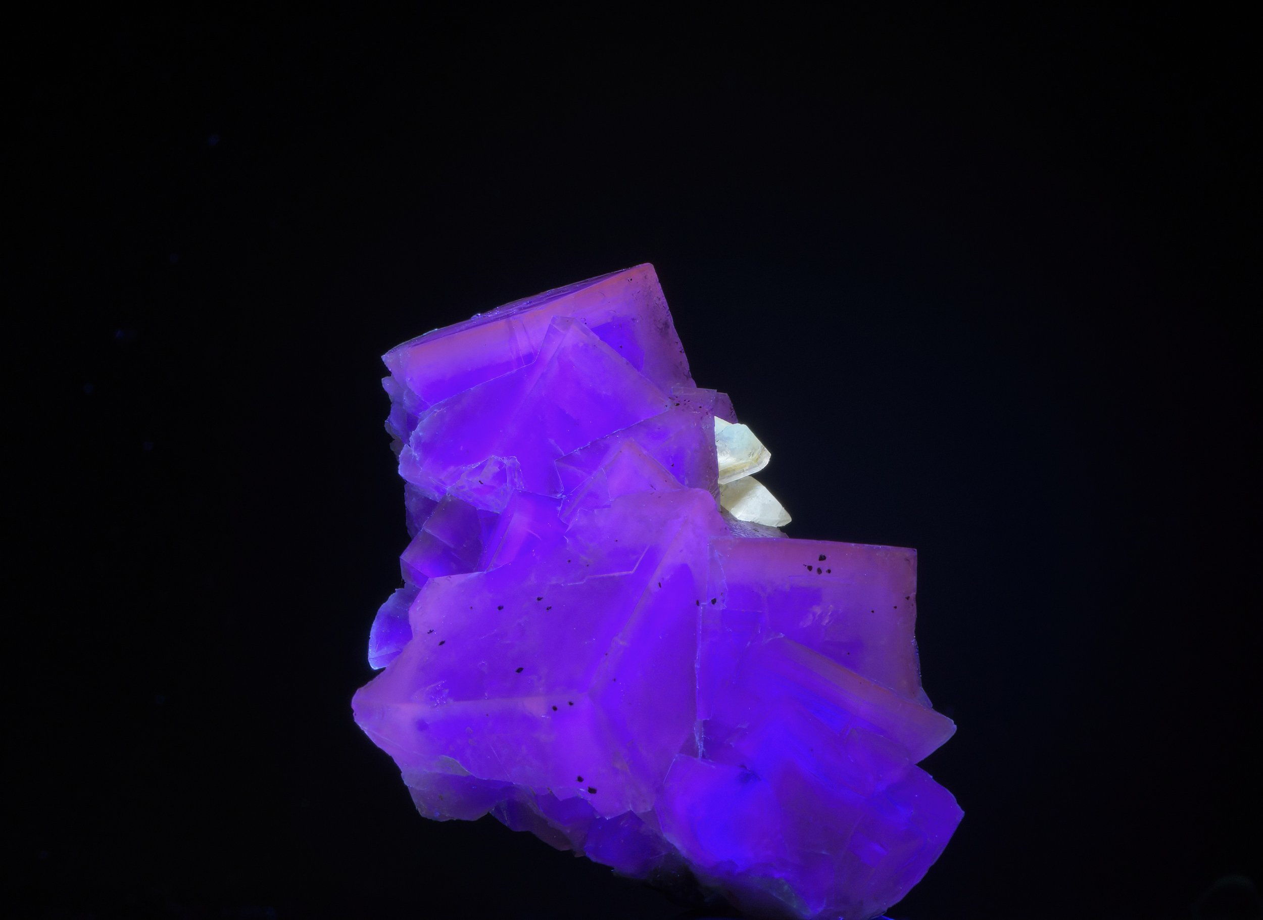
The central Mexican stem succulent, Echeveria bifida, shown illuminated at three light spectra. From left to right, daylight (535 nm), longwave UV (365 nm), and midwave UV (310 nm). Because many echeverias are prized for their attractive wax blooms, it is interesting to see that this species does not fluoresce as might be expected based on observations of other species in the genus (i.e., pale blue), under longwave UV. The center image clearly illustrates that, despite being a plant native to both desert and Tropical Dry Forest ecosystems, it has dispensed with a heavy cuticular wax coating on its stem and leaves leaving its chlorophyll-rich above ground tissue mostly unmasked. Author’s plant. Images: ©Jay Vannini 2023.
Other plant pigments fluoresce somewhat differently, with anthocyanins (an important subgroup of flavonoid pigments) fluorescing orange under midwave UV; carotenoids fluorescing light green under longwave UV (Kleinegris et al., 2010); and betalains also fluorescing greenish yellow to green in plants belonging to the Caryophyllales (Guerrero Rubio et al., 2020). Autofluorescence of some lesser known plant pigment groups has been known since the 1980s, although only recently documented in detail (Gandía-Herrero et al., 2005). The dominance of one or more of these plant pigments, coupled with an immense variety of epidermal structures and coatings that have evolved among the world’s flora, results in a surprising range of colors being reflected in longwave UV-fluoresced tropical plants as documented in this article.

My admittedly superficial examination of UVIVF in tropical ferns so far has revealed several – to my mind – astonishing examples of multi-chromatic longwave fluorescence in both terrestrial and epiphytic species. Above, an apparently undescribed dwarf species of Danaea (Marattiaceae) from the cloud forests of Veraguas, Panamá. Besides exhibiting beautiful longwave fluorescence, under daylight this species showcases very striking, blue-green multilayer iridescent pinnae. This family is considered among the most primitive of all ferns and is extensively represented in the fossil record. Author’s plant. Image: ©Jay Vannini 2023.
So far, so good?
Monitoring the intensity of chlorophyll fluorescence (CF) is now used to gauge photosynthetic activity in nature, field, and greenhouse. Techniques to measure post-harvest stress and senescence in fresh produce are measured via a commercial chlorophyll fluorometer such as a FluorPen (Photons Systems Instruments). Pollen, anthers, staminodes, and stigmas of many plants are also autofluorescent, usually reflecting pale blue under UV, and the compounds involved in fluorescence of reproductive parts are derivative of hydroxycinnamoyl. Several recent controlled field studies have shown that bees, including honeybees (Apis mellifera), detect fluorescence in the bluest photon range and are attracted to it, especially at the lower end of the visible light spectrum in the 430 to 480 nanometer wavelengths (Ostroverkova et al. 2018; Mori et al., 2018).
Pale blue-spectra fluorescence of reproductive parts is likely a late-emerging ancestral trait in both monocot and dicot angiosperms (flowering plants) due to their accelerating co-evolution with bee and fly pollinators, likely dating back at least 125 million years ago (Cappellari et al., 2013). In many insects, sub-400 nm wavelength UV perception (i.e., at deep violet and ultraviolet blue wavelengths) is the rule, not the exception.
Most humans have trichromatic vision, derived from three types of light-sensitive cone photopigments in their retinas. Color blind individuals aside, humans and some other mammals see colors as some combination of blue, green, and red as a product of specific wavelength receptors in these cone cells. That said, color perception is also influenced by light intensity in fore- and backgrounds and viewing colors of any given object against any other than dark surroundings (i.e., in the so-called “aperture mode”) will likely distort the viewers’ percept (Lee, 2008). Likewise, as light levels decrease, luminance sensitivity in human eyes shifts towards the blue end of the spectrum. This phenomenon is known as the Purkinje Effect and is readily observed in garden and household settings when red flowers appear ever bluer under natural light as evening falls.
In rare cases, some humans – invariably women – possess an inherited genetic mutation that is associated with the presence of four photoreceptor cell types in their retinas. These individuals are now referred to as tetrachromats (i.e., possessing four types of cone cells), and a small subset of these individuals apparently do have exceptional color vision (Jordan & Mollon, 2019). Studies of these rare, color-perception “gifted” individuals’ observations are valuable in understanding how other diurnal tetrachromatic vertebrates may see the world around them, especially in response to UV light. People who lack lenses in one or both of their eyes, a condition known as aphakia, are also able to see UV light as whitish blue to whitish violet.
Many other animals, including examples in all higher vertebrate classes, also possess color vision that perceives UV well into the upper boundary of the midwave range (Jacobs, 2013). It is now known to be far more prevalent than previously believed (Withgott, 2000). Birds, well-documented at this point to have tetrachromatic vision, are one good example of this. Hummingbirds were first shown to perceive lower wavelength UV-B in the late 1970s (Goldsmith, 1980), showing the importance of floral guides in plants for pollinators other than arthropods. Some frugivorous birds have been shown to be attracted to berries that exhibit UV fluorescence.
Once it became apparent that birds do indeed perceive some colors differently than we do, researchers rushed to museum ranges to examine study skins under UV lights looking for new color markers in plumage among closely related species (Withgott, 2000).
Ultraviolet vision in fish and lizards has also been known for some time (Fleischman et al., 1993; Siebeck et al., 2010; i de Lanuza & Font, 2014). Among other advantages, enhanced UV perception in vertebrates is used to improve foraging success and for to enhance visual signaling in courtship.

Near visible UV light sources emitting between 385 and 395 nm produce extremely vivid electric purple to neon pink reflections in a wide variety of flowers and foliage that exhibit more complex color schemes at lower UV spectra. Here, a group of the ubiquitous and sometimes invasive Lily of the Nile (Agapanthus africanus) under 390 nm illumination. This showy South African amaryllid is a popular bedding plant with landscapers. It has subtle gray and blue colors under 365 nm but has gone fully “Disco Barbie” under near-PAR UV. Image: ©Jay Vannini 2023.
However, it is worth emphasizing here that enhanced UV perception in animals is just that; color vision that encompasses mid- and longwave UV spectra does not translate into their seeing their surroundings as if through some sort of DayGlo filter.

A time exposure of Coconut Palms, Cocos nucifera, on a beach photographed with infrared sensitive film. Image: ©Peter Rockstroh 2023.
Photographers have explored both UV fluorescence and infrared (IR) luminescence in flowers and plants for decades. While mostly outside of the scope of this article, for reference purposes the IR spectrum ranges from just past the edge of visible light at ~700 nm, through to 1 mm. IR filters and film produce interesting visual effects. An image shot with IR film is shown left.
And Onto Plant Fluorescence
After scanning a small sample of tropical ornamental plants for evidence of showy fluorescence under longwave UV-emitting handheld lights, it was immediately apparent that some were reflecting UV in color ranges far more varied and complex than just bright red or pale blue.
Queries made to a few well-informed botanist friends and colleagues revealed that little had been published on the topic. I checked online for signs that a systematic evaluation of fluorescence under longwave UV had been conducted in the past. As far as I have been able to determine, it has not. The few references I was able to glean led me to conclude that curious gardeners and photographers who had illuminated ornamental plants with UV flashlights had decided that a). most if not all lowland rainforest plants exhibited red fluorescence due to their thin leaf cuticles that effectively unmasked chlorophyll, and b). that succulent xerophytes all fluoresced blue due to the presence of epidermal wax coatings. One early adopter who has explored UVIVF and plant parts in his creative photography over the past decade is California-based Craig Burrows, but his focus has primarily been on the flowers of native plants (Yearsley, 2022).
There are thousands of images posted online of common flowers and plant species fluorescing red or blue under longwave UV light depending on their dominant plant pigment and epidermal features. There is an entire subset of images on the internet that are labeled “bee vision” or “butterfly vision” photography. Some of the color combinations shown in these images are based on flawed assumptions as to how insect pollinators view primary colors, so these images should be viewed with that in mind. While on this topic, “bee vision” photographers also omit the distortion that extreme focal convexity (to 300°) and the blurring that bees’ nearsightedness generates.
Many insect pollinators have very sophisticated color vision, and recent work has shown that because of variations in photoreceptor sensitivities, color perception varies even between sexes of the same swallowtail butterfly (Papilio) species, as well as across members of the same insect genera (Chen et al., 2016).
Researchers now know that plants and their floral parts often fluoresce under longwave UV in colors that are attractive to well-known insect pollinators, especially bees, flies, butterflies, moths, and beetles and that areas of notable fluorescence are both differentially-zoned and of varying intensities in many disruptively colored and showy tropical plants.
What has been lacking so far is a systematic look at the far corners of the world’s flora through the filter of UV light from shortwave to near visible longwave spectra, so I thought I would open the conversation here.

Tubercles on the leaves of stem succulents and others provide additional visual treats to UV-lit plants. Shown here, leaf details of a northeastern South African alooid, Gasteria batesiana ‘Barberton’, illuminated with 365 nm wavelength lights under blackout conditions. Note the red reflective, lightly-masked, chlorophyll-rich new growth. Author’s plant. Image: ©Jay Vannini 2023.
The images shown in the article are not intended to illustrate how other animals with color perception ranging well into the UV spectrum might see these plants. Rather, it is to show that many of these plants possess a surprising heterogeneity of colors when illuminated at lower wavelengths not visible in daylight, and that curious plant collectors would be wise to explore the UVIVF phenomenon. That said, our ability to consistently generate UVIVF in many plants is also a valuable insight for researchers, since it may provide clues as to why certain stem and leaf adaptations and colors evolved in certain species.

Not all plants possess showy fluorescence under UV illumination. Some Mexican and South American globular cacti species just glower under longwave UV. Above, a Bolivian Rebutia canigueralii f. rauschii trying hard to muster some violet tones under a 365 nm light source. Author’s plant. Image: ©Jay Vannini 2023.
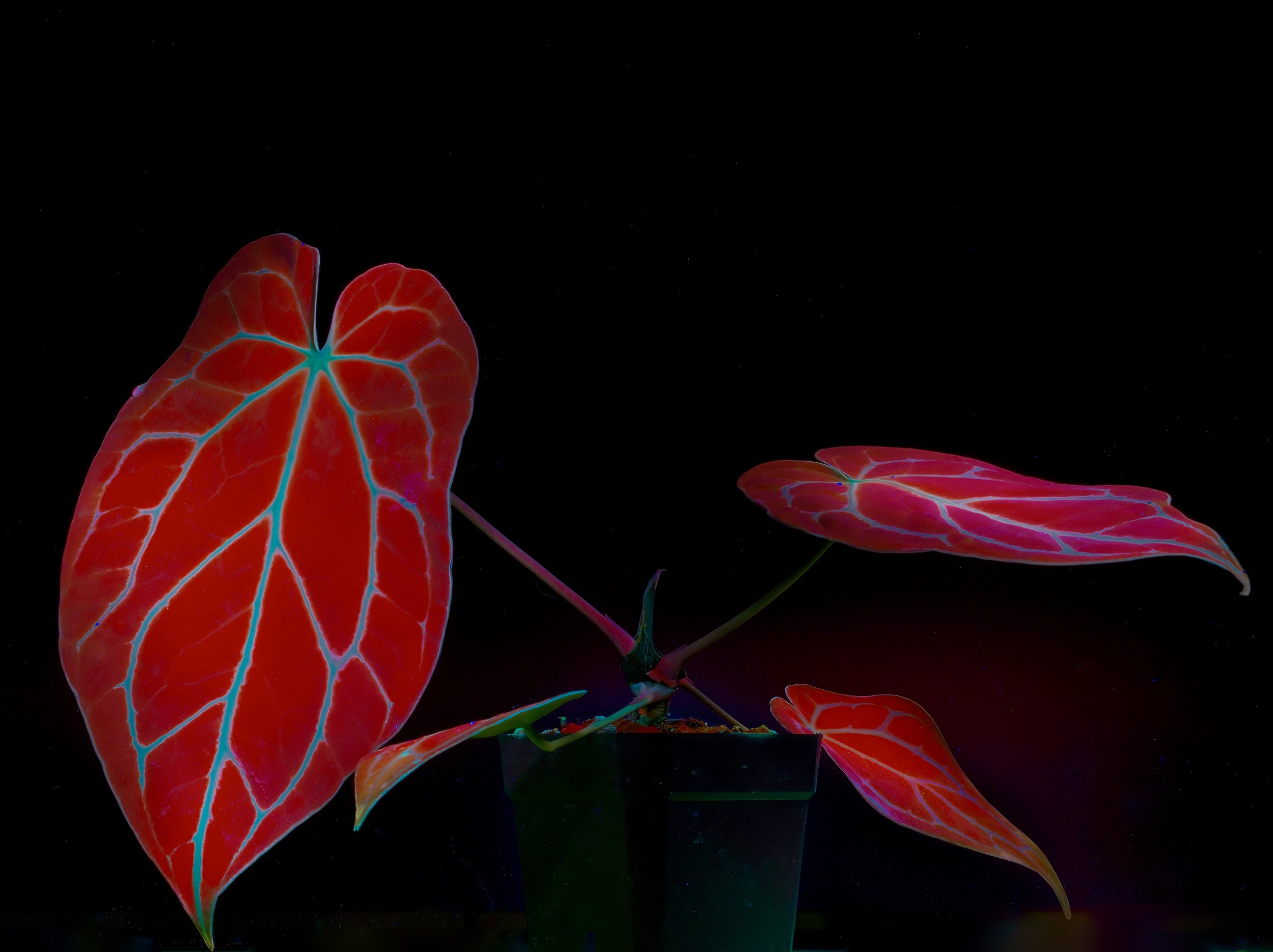
Unsurprisingly, four megadiversity plant genera that are popular in ornamental horticulture – Anthurium (Araceae), Bulbophyllum (Orchidaceae), Euphorbia (Euphorbiaceae), and Begonia (Begoniaceae) – include many species that exhibit showy, multicolored fluorescence (pers. obs.). The elaborate fluorescence models exhibited by some members of the first three genera can be surprisingly heterogeneous even across what we consider closely related species. Conversely, many begonia leaves, even those with bold contrast colored patterns, generally fluoresce some intense shade of concolorous red under longwave UV, although there are some notable outliers to that general rule.
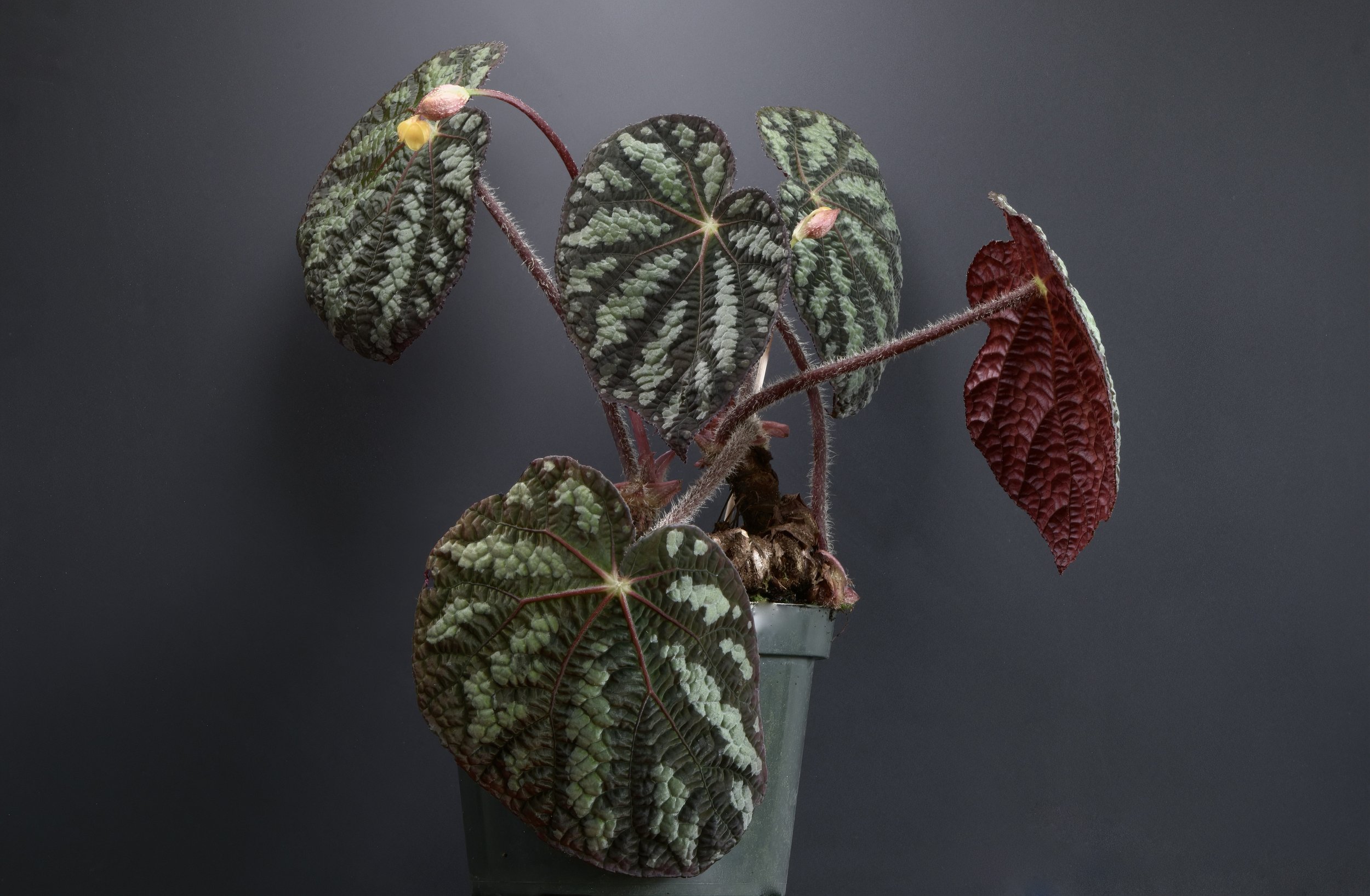
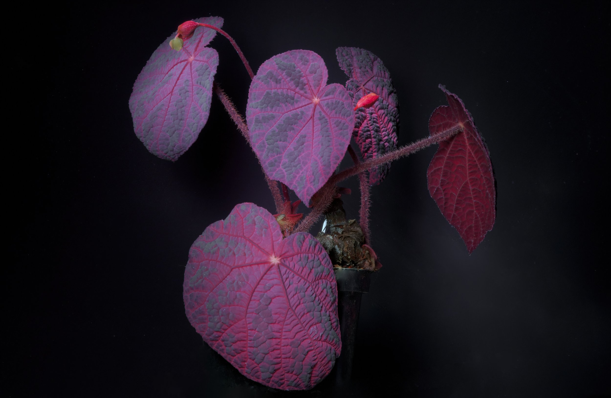
For every rule there always seem to be exceptions. This one’s definitely worth a look. Most Begonia species that I have examined under longwave UV fluoresce bright red under blackout conditions. Here is one remarkable exception to that finding, the subtropical B. xanthina f. pictifolia from Assam State in northeastern India. Multiple plant pigments are on display that reflect quite differently both in daylight and under 365 nm illumination. The visible pale blue iridescence is accentuated by a bullate leaf texture and a dense coat of very short, light reflecting trichomes on their leaves. Note that the dark red lower surface on the leaf on the right of both images does not fluoresce. Under midwave UV at 310 nm, the foliage on this plant fluoresces a uniform metallic blue with light pink primary veins. Author’s plant. Image: ©Jay Vannini 2023.
Karabourniotis et al. (2016) rightly point out that a plant’s epidermis is a chloroplast-free “selective optical filter”. Environmentally adapted epidermal features on stems, leaves, and floral parts such as papillose cells, tubercles, crystalloid/epicuticular waxes, cuticle thicknesses, presence of surface glands, helicoidal cells, etc. all play an important role in modulating light capture and influencing the nature and intensity of UV fluorescence in any given plant. Papillose leaf surfaces, along with those having elongated, multicellular trichomes and heavy coatings of cuticular waxes reveal extremely irregular micro-relief when examined under high magnification, can be highly reflective, and serve as effective water repellents even when the leaves are submersed for days (Pierce et al., 2001; Karabourniotis et al., 2021).
Plants are extremely sensitive to the intensity, wavelength, and distribution of spectral light in their environment. Recent studies have shown that high levels of exposure to midwave UV (UV-B) can produce either positive or negative physiological responses in plants depending on the species. Lower levels of exposure to midwave UV radiation can prompt the production of photoprotective chemicals in leaf tissue. One such response involves the accumulation of flavonoid pigments in the upper layers of the epidermis and in chloroplasts where they effectively function as UV-blocking sunscreen (Palma et al., 2021). These concentrations of flavonoids, especially anthocyanin, may partly explain the prevalence of red and orange UV reflectance in some xerophytes that would be expected to exhibit cuticular wax coatings and blue fluorescence but do not (pers. obs.).
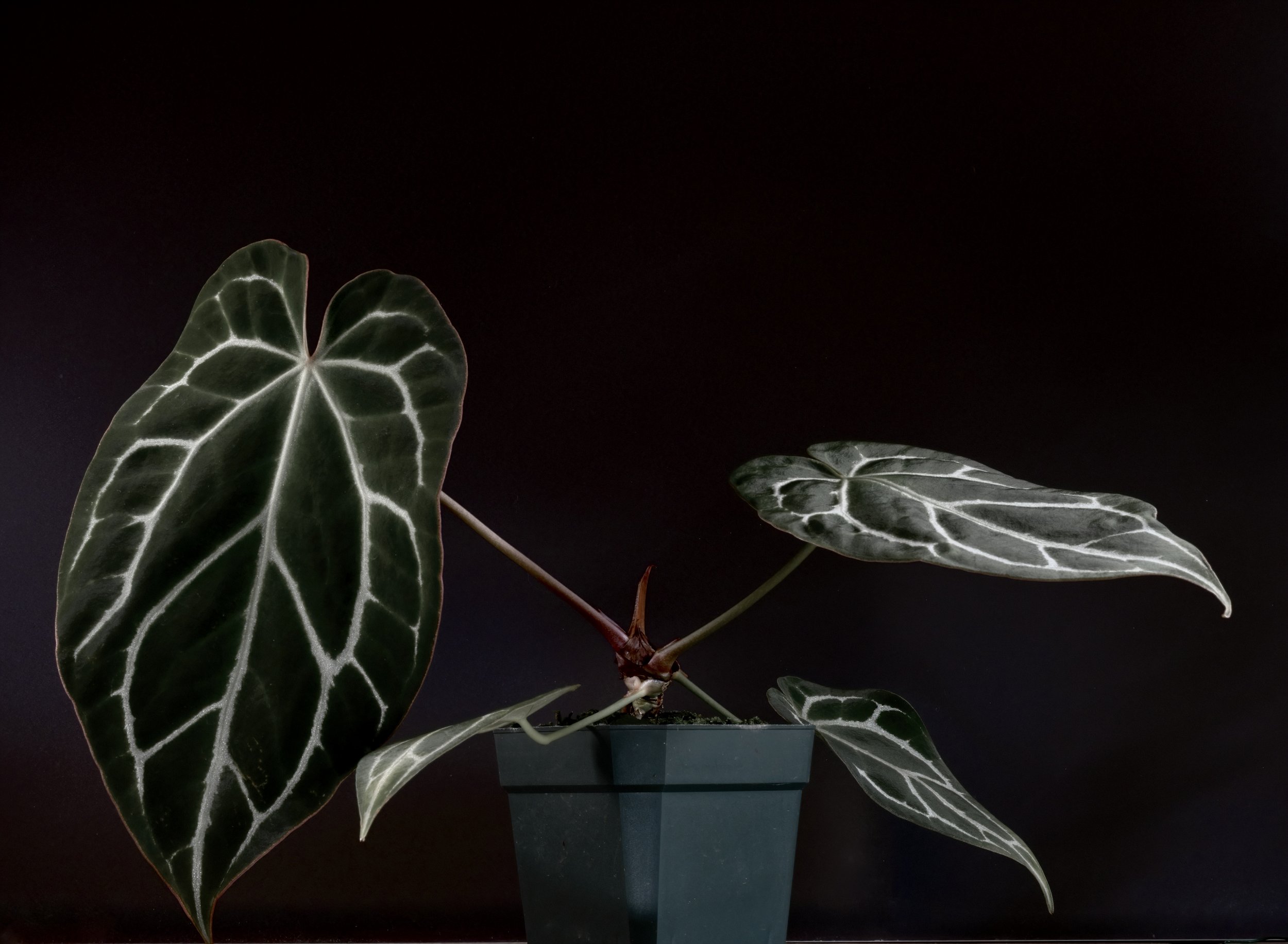
Some Anthurium species originating from heavily-shaded understory niches in Panamanian and Colombian rainforests possess some of the most intense leaf fluorescence observed in the Araceae. Their dark velvety leaves have evolved leaf surface cells that optimize the capture and focus of stray light flecks that penetrate the canopy and onto the forest floor. In person, the reflected red is deeply saturated and seemingly three dimensional. Ultraviolet blue contrast primary veins are a ubiquitous feature in “black velvet” anthuriums and, together with chemical lures emitted by their flowers, may play a role as visual guides when attracting short-sighted euglossine bee pollinators. Shown here, a young, artificially propagated Anthurium crystallinum ex-founders that originated from Colombia’s Antioquia Department. Author’s plant. Images: ©Jay Vannini 2023
Super-saturated candle apple red fluorescence often enhanced by a nacreous sheen is apparently restricted to plants with velvety leaf surfaces that are illuminated with longwave UV. I observed this phenomenon in a number of Anthurium, Philodendron, and Begonia species known to possess lens-shaped epidermal cells (Karabourniotis et al. 2021; pers. obs.). There is a strong correlation between the presence of papillose epidermal cells on the upper leaf surfaces of these plants a distinctive “metallic” luster to their fluorescence under longwave UV. This is likely explained by the ~180° x 360° reflectance that one would expect to observe in extreme examples of convex papillose cells on the upper leaf surfaces of understory plants adapted to very dark microhabitats.
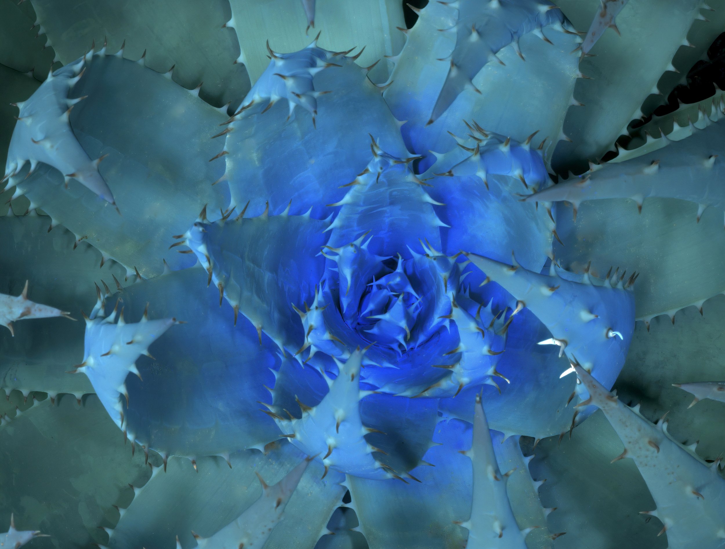
A look-down on Aloe peglerae lit with 365 nm wavelength UV. This endangered xerophyte from North West and Gauteng Provinces in South Africa displays a noticeable glaucous bloom on its leaves in daylight from cuticular waxes. The image shows a particularly vivid example of the type of blue fluorescence observed in many stem succulents. Author’s plant. Image: ©Jay Vannini 2023.
Taken as a group, cuticular waxes are long-chain fatty acids and derivatives that cover stem, leaf, and some floral surfaces such as peduncles and bracts. They are complex mixes including aldehydes, alkanes, primary alcohols and, in some species, triterpenoids. Their composition varies between species, occasionally even between different ecotypes of the same taxon, the developmental stage of the leaf, and on which side of the leaf they occur (Ahmad et al., 2015; Simões et. al., 2020).
Because the chemical composition of cuticular wax compounds can vary across both leaf surfaces, conspicuously different fluorescence coloration are often evident between upper (adaxial) and lower (abaxial) leaf surfaces (pers. obs.) under longwave illumination. Leaves should routinely be inspected for this contrast when evaluating the presence of UVIVF.

Several central African rainforest origin parrot beak Impatiens species may be the most colorful and intensely reflective of all longwave UV-fluorescent cultivated plants. The image shows leaf details of I. cf. clavicalcar from the Democratic Republic of Congo. At least two species, including this one, reflect up to five colors at 365 nm – mauve, blue, green, yellow, and red + white – on the same individual’s stems, leaves, and emerging flowers. They are excellent studies of the complex interplay of plant pigments, zoned epicuticular waxes, epidermal cell structures, and response to changing light angles under UV lights. Author’s plant. Image: ©Jay Vannini 2023.
Hand-held UV light sources are now available at accessible prices and at several wavelengths ranging from ~250 nm through to 395 nm. Dedicated flash units are also available that can be set up for both infrared and UV photography. See the Kolari KV FL-1 UVIR flash unit for one example, with UV emitted at 345-380 nm (via a UV bypass filter) and IR illumination projected at an 850 nm wavelength.
It is important to note that light leak from the margins of low-end lamps as well as stray light from ambient sources will distort colors in UV photography. I use the “can’t-see-my-hand-right-in-front-of-my-face” rule of thumb in terms of determining adequate blackout levels when shooting UV-luminated images. Depending on the amount of light pollution infiltrating the studio setting, the wavelength of the UV emitting source, the proximity of the light source to the subject, and the intensity of the subject’s fluorescence, plant parts may not reflect their true UVIVF colors.
The topic of use or need for UV filters for DSLR lenses is a complex one and beyond the scope of this article. Likewise, the topic of UV Reflectance Photography, which uses different equipment and produces quite different results to those shown here, is one for others to illustrate. Those interested in the science and debate over UV filters for modern lenses, and the arcana of UV Reflectance Photography will find a wealth of information at the UltravioletPhotography.com website linked in references. For what it’s worth, I do use UV filters on my lenses, primarily for lens safety.
For comparative purposes that are useful to photographers, Wien’s displacement law for conversion of color temperature measured in Kelvin (K) to peak wavelength to nanometer equivalency is 3,000,000/K value. So, a daylight temperature light source such as one adjusted to 5,600 K is equivalent to a ~535 nm wavelength.
I will re-emphasize this point at the end of the article but take this opportunity to clarify the risks to misusing UV-C and UV-B lamps such as those used to produce some of the images shown here. These tools, together with more readily available longwave UV-A flashes, should always be used with UV-blocking eyewear, should never be pointed at your own, others’, nor any animals’ eyes. Both temporary and long-term damage to corneas, retinas, and exposed skin may result from their improper use. Mishandling or overexposure can cause ultraviolet keratitis, a distressing and extremely painful condition. Long exposures using UV-C also generate hazardous ozone emissions, so these sessions should be conducted in well-ventilated spaces. The lamps should be stored – preferably with their batteries or power sources removed – where they cannot be accessed by children, adolescents, and curious/stupid adults. UV-C lamps should also not be pointed at exposed skin surfaces.
Please remember that UV damage to eyes is cumulative! Do not fool about with high intensity UV-emitting light sources.
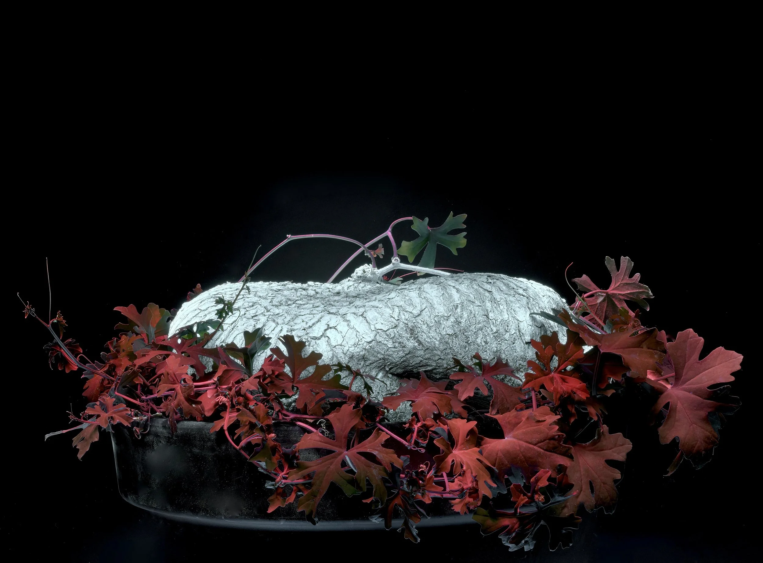
The Slimlobe Globeberry, Ibervillea tenuisecta, is a Chihuahuan desert geophytic gourd that is prized in cultivation for its large, flat, pancake shaped tubers. Subterranean in nature, they are usually grown with the tuber exposed as an ornamental caudiciform. Despite inhabiting harsh, arid conditions and sometimes growing fully exposed to to almost constant sunshine, this species has foregone cuticular waxes to protect its leaves and instead appears to rely on flavonoid pigments in its foliage to act as midwave UV-blocking sunscreen. New Mexico and Texas, USA to San Luís Potosí, México. Author’s plant. Image: ©Jay Vannini 2023.
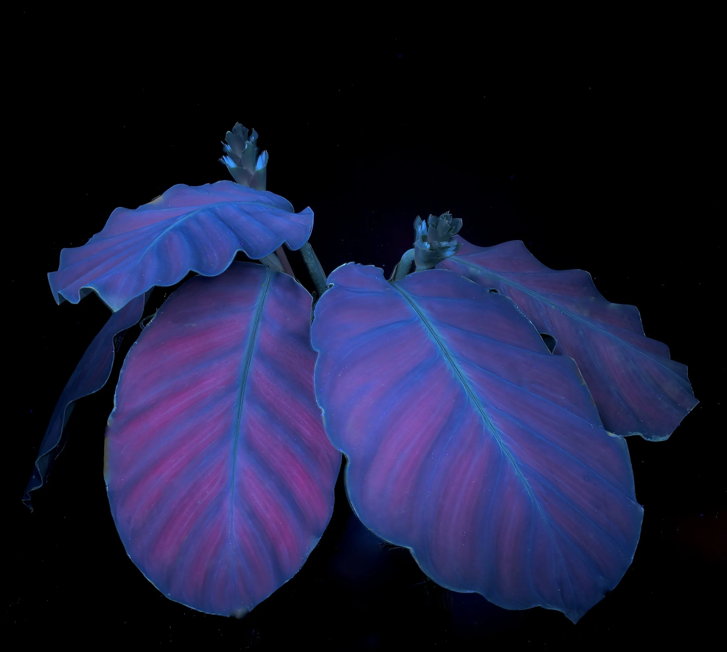
Calatheas and other tropical arrowroots can exhibit a wide range of very showy fluorescence color schemes when lit by longwave UV. This is Goeppertia dressleri, a shade-dependent rainforest floor denizen from eastern Panamá and adjacent Colombia. Author’s plant. Image: ©Jay Vannini 2023.
Aloe species and related genera are extremely variable with regard to color reflectance when illuminated with long- and midwave UV under blackout conditions. Above, the handsome alooid Haworthiopsis coarctata var. adelaidensis, a crevice dweller on rocky outcrops in savanna habitats in South Cape Province, South Africa. It’s sheltered origins may explain why it has dispensed with a heavy cuticular wax coating on stems and leaves. Author’s plant. Image: ©Jay Vannini 2023.

A mature Ridley’s Staghorn Fern, Platycerium ridleyi, from eastern Myanmar fluorescing under 365 nm light. Many tropical fern species that are popular in cultivation exhibit showy, polychromatic fluorescence. This handsome Indomalayan fern is a high rainforest canopy epiphyte and the conspicuous cuticular wax coating evident on both shield and fertile fronds likely provide substantial protection from the effects of excess sunlight. Author’s plant. Image: ©Jay Vannini 2023.

The flowers of many ornamental amaryllids that are native to the northwestern Andes have stunning longwave fluorescence under blackout. Above, Eucrosia euchrosioides from seasonally dry ecosystems on the Pacific slope of southwestern Ecuador and northwestern Perú. Author’s plant. Image: ©Jay Vannini 2023.
Gallery – Representative Plant Families
This is by no means intended to be taken as an exhaustive examination of UVIVF in cultivated tropical ornamental plants, but rather is intended to be used as a first draft basic roadmap for collectors and researchers to explore this field further.
There are 118 images included below of colorful UV-fluorescent tropical plant species representing 40 genera and 26 families.
I have included brief notes on some of the better known, mostly subtropical and tropical plant families that exhibit colorful fluorescence under 365 nm wavelength UV light, together with photographs of them under this type of UV illumination. Plants were subjectively sampled – based on what I determined were promising leaf or floral characters – from my personal collection in California which includes ~1,700 species and proprietary hybrids from 56 currently recognized plant families. Because these images were captured over the eight week period leading up to publication of the article, many flowering or semi-dormant plants I would have liked to include were not in a condition to illustrate their more interesting features…
To continue reading the rest of this and other Premium Content articles, please subscribe to Esotérica Exclusiva
Follow us on:
Access to deeper dives and behind the scenes looks into advanced tropical horticultural practices as well as observations on fieldwork, plant collection management, and Neotropical wildlife.


